The heart is one of the most vital organs in the human body and is crucial in sustaining life. Research in cardiac medicine has consistently been one of the most complex and important areas in the medical field, and thanks to the adoption of 3D scanning technology, new research is now possible.
Such as the research by Dr. Rohit Loomba, a pediatric cardiac physician at Lurie Children's Hospital in Chicago, is conducting research using 3D scanning technology to perform detailed heart scans. Drawing on his clinical experience, he refines these scans into a collection of heart specimens. This work provides accurate models for academic research and enables him to share his scanning results with colleagues and the public through social media.
This article explores how Dr. Loomba uses Revopoint's MINI2 and MIRACO 3D scanners to create detailed heart specimen scans, expanding possibilities for medical research.

Studying heart disease requires a clear understanding of complex anatomical structures. While traditional 2D images and anatomical specimens are useful, they often fall short of representing these intricate 3D forms. So, 3D scanning improves analysis accuracy by creating accurate digital models that allow researchers to examine details from various angles.
Especially in pediatric cardiology, the research subjects are usually newborns' and infants' small, delicate, and complex hearts. This necessitates high standards for scanning equipment in terms of resolution, speed, and usability.
MINI 2’s First Heart Scan

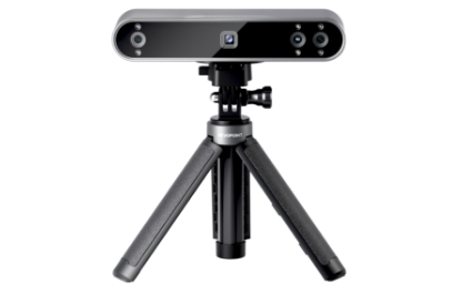
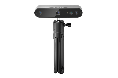
Dr. Loomba first used the Revopoint MINI 2 scanner to scan heart specimens. This binocular blue light 3D scanner is designed for high accuracy, especially in capturing fine details of small objects.
In a LinkedIn post, Dr. Loomba shared his experience scanning a heart specimen with the MINI 2 3D scanner, which had undergone Baffe’s surgery. He wrote in the post:
Heart w/ transposition and Baffe’s procedure. IVC baffled to left atrium and the right pulmonary veins to the right atrium! Click to check out the 3D scan and model of a specimen in our collection! Scanned with the Revopoint 3D Mini 2!

(Case image from https://www.linkedin.com/posts/rohit-loomba-69266824a_transposition-sp-baffes-i-133-activity-7216258125434167296-TqnR?utm_source=share&utm_medium=member_desktop)
This scan showcases the impressive resolution of the MINI 2, capturing intricate structures in the specimen. This detail supports further research and allows professionals to examine valuable medical models closely, contributing to medical development and education.
MIRACO Does Even Better

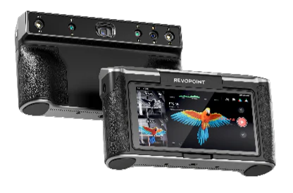
With his research deepening, Dr. Loomba decided to try another 3D scanner, this time the Revopoint MIRACO, a standalone 3D scanner. This scanner combines scanning and post-processing with a clear 6-inch 2K backlit touch screen that is adjustable up to 180° for easy viewing. It's especially handy for larger heart specimens.
Standalone Capabilities
Dr. Loomba can scan with MIRACO without needing a laptop. This is a significant convenience for those working in a laboratory environment with chemicals such as formalin.
Capturing Fine Details
Dr. Loomba was impressed by its scanning capabilities. When scanning a baby’s heart, about the size of a fist, MIRACO produced a clear and detailed model, capturing its fine structures despite the small size.
Dr. Loomba plans to explore MIRACO's capabilities and optimize the scanning process to enhance medical research and technology integration. His work demonstrates the applications of 3D scanners, such as the Revopoint MINI 2 and the MIRACO, as valuable tools for advancing cardiology research.

3D scanning technology is not just an addition to traditional research methods. It also plays a significant role in advancing medical visualization and data. For professionals like Dr. Loomba, these tools improve research efficiency and offer valuable teaching resources. As technology evolves, the use of 3D scanning in medicine will continue to expand.
We look forward to more practitioners using innovative technology in research and teaching to tackle complex medical problems and bring hope to patients.













Leave a comment
This site is protected by hCaptcha and the hCaptcha Privacy Policy and Terms of Service apply.